Fig. 3.
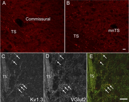
Confocal images of the NTS labeled with Kv1.3. A: confocal image of a horizontal NTS section in the commissural region. Fine fibers in the solitary tract (TS) exit the tract and innervate neurons medial to the tract. B: confocal image of a horizontal section of the NTS showing the medial nucleus of the NTS (mNTS), the site of termination of baroreceptor axons. Immunostaining of both sections with anti-Kv1.3 antibodies (red) showed the presence of the protein in the fibers in the tract and in the neuropile but not in cell bodies. Images are stacks of 12–15 z-sections taken at 0.3-μm intervals. Scale bars = 15 μm. C–E: confocal images of anti-Kv1.3 and anti-vesicular glutamate transporter 2 (vGlut2) in the TS and medial NTS. C: anti-Kv1.3 was present in the afferent sensory fiber tract (TS) and in discrete bouton-like structures (arrows). D: vGlut2 immunoreactivity was present in the same structures. E: merged image of Kv1.3 (red) and vGlut2 (green). Each image is a stack of four z-sections. Scale bars = 20 μm. Similar anti-Kv1.3 labeling was obtained in the NTS from four other animals.

