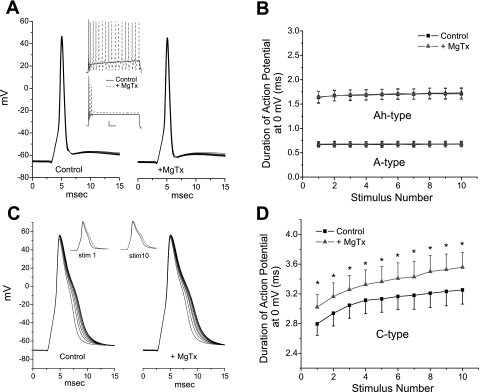
Fig. 5.
Effects of MgTx on neuronal discharge. A: example of an A-type neuron (Na+ current blocked by TTX) responding to 10 brief (0.35–1.5 ms) stimuli delivered at 20 Hz. There was no change in the duration of the action potential with frequency, nor did MgTx (1 nM) have an effect on duration. However, a longer-duration stimulus (top inset) increased discharge from one action potential in the control solution (solid black line) to repetitive firing in the presence of MgTx (dashed red line) or increased the amplitude of the depolarization to the constant current stimulus (bottom inset). Scale bars in insets = 50 ms, 10 mV. B: mean duration of the action potential at 0 mV plotted for 3 A-type neurons for each of the 10 stimuli in control solution and in the presence of MgTx. A group of six Ah-type neurons also did not respond MgTx. C: MgTx broadened the C-type action potential but did not eliminate the stimulation-dependent lengthening of the action potential (20-Hz stimulation), as shown in control solution and in the presence of 1.0 nM MgTx. Left inset, the first action potential in the 20-Hz train in the control solution (black) compared with the first action potential in the presence of MgTx (red); right inset, the 10th (last) action potential of the series without (black) and with (red) MgTx. D: mean duration of the action potential of eight C-types neurons in the absence and presence of MgTx plotted for each of the 10 stimuli. Four of these neurons had been previously incubated in α-dendrotoxin to eliminate any effect of MgTx on Kv1.1, Kv1.2, or Kv1.6. *P < 0.05 (paired t-test).

