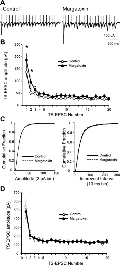
Fig. 6.
MgTx augments TS-evoked excitatory postsynaptic currents (EPSCs). A: representative tracings of TS evoked EPSCs that were recorded under control conditions (in artficial cerebrospinal fluid) and after MgTx (20 nM). The TS was stimulated at 20 Hz. Note the increase in the TS-EPSC amplitude, especially the first event. Shown is an average of five current sweeps. B: synaptic events were grouped according to their initial current amplitude. Data shown are the mean TS-EPSC amplitudes for 20 events whose initial amplitudes were <300 pA during control and MgTx application. n = 15. *P < 0.05 (two-way repeated-measures ANOVA). MgTx elevated EPSCs primarily at the beginning of the stimulus train. C: spontaneous EPSC frequency was also elevated in MgTx-sensitive currents. The cumulative probability of sEPSC amplitude distribution (2-pA bin, left) was not altered in MgTx. On the other hand, the cumulative fraction of spontaneous EPSC interevent intervals (10-ms bin) illustrated a small leftward shift in the presence of MgTx (right). The analysis was performed on the entire sample of events. D: mean TS-EPSC amplitude for 20 events during control and MgTx application for three cells whose initial TS-EPSCs were >300 pA and did not respond to MgTx application. Note that the amplitudes of the EPSCs that were sensitive to MgTx (B) were significantly smaller than those of MgTx-insensitive NTS neurons.

