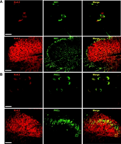
Fig. 8.
Colocalization of Kv4.2 and neurokinin 1 (NK1) or PKCγ. A: representative confocal images of Kv4.2 (red) and NK1 (green) immunofluorescence in dorsal horn neurons and in the mouse dorsal horn. B: representative confocal images of Kv4.2 (red) and PKCγ (green) immunofluorescence in dorsal horn neurons and in the mouse dorsal horn. Merge of Kv4.2 with NK1 or PKCγ is shown in yellow. Original scale bars: cultured cells, 40 μm; slices, 200 μm.

