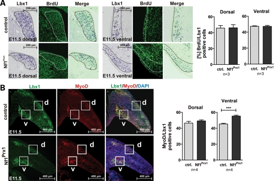
Figure 4.
Muscle progenitor proliferation and myoblast differentiation in Nf1Prx1 mice at E11.5. (A) Co-labeling for Lbx1 (in situ hybridization) and BrdU (antibody staining) on sections of E11.5 embryos. Proliferation index was assessed by bromodeoxyuridine (BrdU) labeling for 1h prior to sacrifice. Quantification of cells in the encircled areas is shown right for dorsal and ventral muscle masses, respectively. No differences in proliferation of Lbx1-positive cells were detected in Nf1Prx1 mutants. (B) Immunodetection of Lbx1 (green) and MyoD (red) expressing cells on the transverse sections of the forelimb buds identified migrating muscle progenitor cells and myoblasts, respectively. (B) Quantification of myoblast differentiation assessed by the relation of MyoD- versus Lbx1-positive cells. The ratio of MyoD-positive to Lbx1-positive cells is increased in the ventral region in mutant mice, which is not caused by the decrease in Lbx1-positive cells but rather by the more abundant MyoD-positive cells (data not shown). The increase appears to reflect a premature induction of the myogenic differentiation of the migrating progenitor cells in the ventral limb bud region.

