Figure 7.
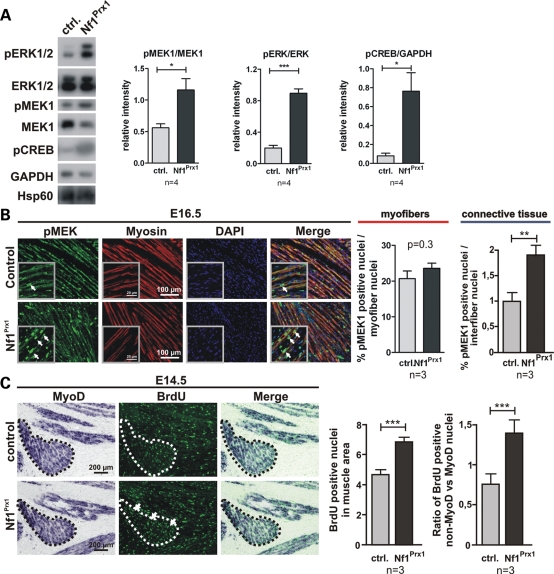
Hyper-activation of Ras signaling in the Nf1-deficient muscles. (A) Western blot analysis of MAPK signaling in adult mice (P90). MEK1, ERK1/2 and CREB are hyperphosphorylated in the mutant muscles. Blots probed with antibodies against pMEK1, pERK1/2 and pCREB were quantified densitometricaly (right). (B) Immunohistochemical detection of pMEK1 on longitudinal sections of the triceps muscle at E16.5. Magnified area is shown in the inserts. A robust pMEK1 signal was detected in the Nf1Prx1 muscles in the myosin-stained myofibers as well as in muscle connective tissue cells (arrows). Quantification of the MEK1-positive nuclei in the myofibers and interfiber space (right panel). WT values were set as 1. (C) Combined in situ hybridization for MyoD and immunolabeling for BrdU (1 h pulse) showing increased proliferation (BrdU, green) of cells in the MyoD-positive area (dotted line) at E14.5 (right panel). This was carried mainly by non-MyoD-positive connective tissue cells, which showed a higher proliferation index than MyoD-positive myoblasts (right).

