
Figure 7.
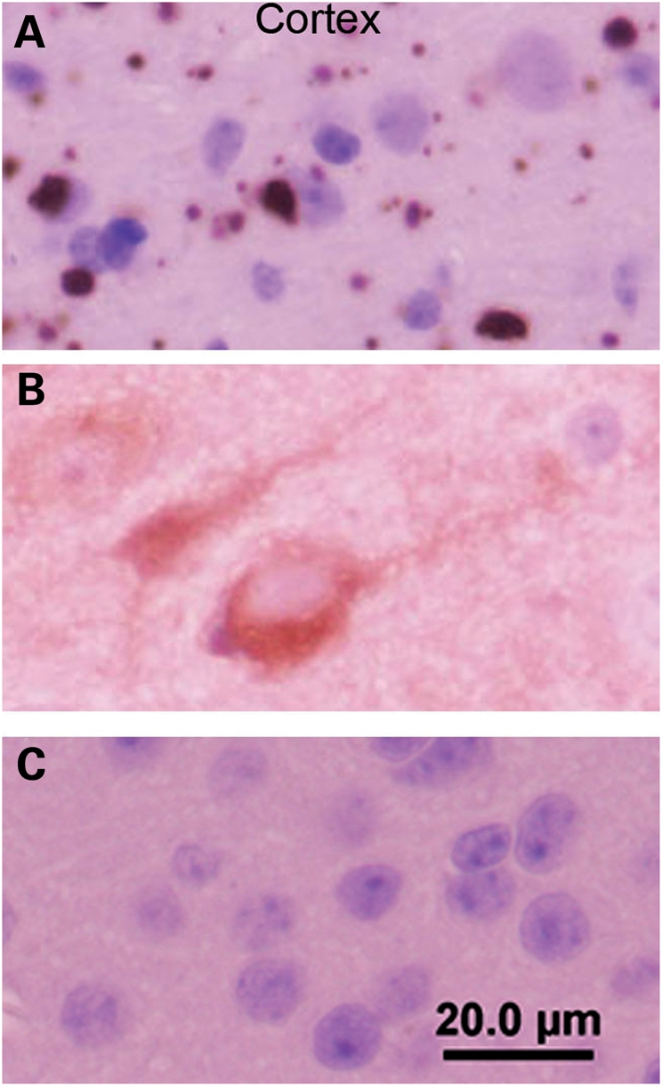
Cortical inclusions are predominantly cytoplasmic in N586-82Q-C63 mice. End-stage line N586-82Q-C63 mice (8 months old) exhibit punctate immunostaining in the neuropil of the cortex with larger perinuclear aggregates also visible (A). In contrast, age-matched N586-23Q-A2 mice exhibit diffuse staining of the cell bodies of a subset of cortical neurons (B). Age-matched NTg littermates of line C63 mice exhibit very low levels of htt immunoreactivity when stained and processed in parallel (C). These images are representative of what was visualized in three different symptomatic animals on at least three tissue sections from each animal immunostained with the 2B4 antibody (see Materials and Methods).