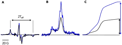
Figure 5. Dynamics of the labeled Tm in the muscle fiber.
A. Overlay of the cw-EPR spectra of labeled Tm in ghost fiber (black) and labeled Tm cross-linked to DITC glass beads in solution (blue). The double arrows correspond to the hyperfine splitting (2 Teff) of the cw spectra. B. Overlay of the ST-EPR spectra of labeled Tm in ghost fiber (black) and labeled Tm cross-linked to DITC glass beads in solution (blue). C. Comparison of the normalized first integral of the ST-EPR spectrum (V'2) of labeled Tm in ghost fiber (black) and labeled Tm cross-linked to DITC-glass beads (blue). The double arrows correspond to the height of the first integral of the normalized V'2 spectra.

