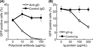
Figure 3.

Involvement of equine herpesvirus-1 (EHV-1) glycoprotein D (gD) in equine major histocompatibility complex (MHC) class I-mediated viral entry. (A) Inhibition of EHV-1 entry by anti-gD polyclonal antibody. EHV-1 Ab4-GFP at a final MOI of 5 was incubated with anti-gD polyclonal antibody (open circles) or control rabbit IgG (solid squares) for 30 min, and the virus–antibody mixture was added to 3T3-A68 cells. After incubation for 2 h, extracellular virus was inactivated by treatment with citrate buffer. Green fluorescent protein (GFP)-positive cells were counted by flow cytometry. (B) Inhibition of EHV-1 entry by gD-Ig fusion protein. 3T3-A68 cells were incubated with gD-Ig (open circles) or control-Ig (solid squares) for 30 min and infected with the EHV-1 Ab4-GFP at an MOI of 5. After incubation for 2 h, extracellular virus was inactivated by treatment with citrate buffer. GFP-positive cells were counted by flow cytometry. Error bars represent standard deviations of three independent samples.
