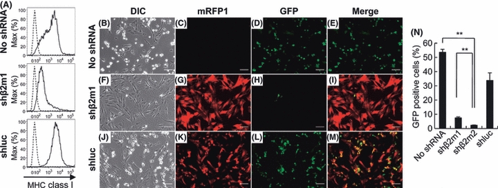
Figure 6.

Knock-down of the cell surface expression of major histocompatibility complex (MHC) class I. E. Derm cells were infected with recombinant lentiviruses carrying both shRNA and mRFP1 expression cassettes. E. Derm cells were transduced with equine β2m-specific shRNA (shβ2m1 and shβ2m2) or luciferase-specific shRNA (shluc). (A) Flow cytometric analysis of the cell surface expression of MHC class I. Cells transduced with shRNAs (solid lines) or nontreated E. Derm cells (dashed line) were stained with anti-MHC class I antibody PT85A. (B–M) Effect of shRNA transduction on the susceptibility of E. Derm cells to equine herpesvirus-1 (EHV-1). Cells were infected with Ab4-GFP at an MOI of 1 for 24 h. Transduction of shRNA lentiviral vectors was confirmed by mRFP1 expression and infection of Ab4-GFP was confirmed by green fluorescent protein (GFP) expression under a fluorescence microscope. Scale bars: 100 μm. (N) After infection with Ab4-GFP, GFP-positive cells were counted by flow cytometry. The bars represent the means from three samples, and error bars show standard deviations. Statistical significance was analyzed by Student's t-test and is indicated by an asterisk (**P < 0.01).
