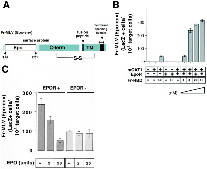
Figure 3.
(A) Schematic diagram of chimeric erythropoietin/MLV envelope protein (Epo-env). The diagram demonstrates the location of erythropoietin inserted in the Fr-MLV envelope protein (Fig. 1B). (B) Rescue of Fr-MLV (Epo-Env) infection by purified RBD. 293 cells or 293-derived cell lines expressing EpoR and/or mCAT1 were treated with a 1:10 dilution of viral supernatant containing Fr-MLV (Epo-env) in the presence or absence of Fr-RBD (40 nM). On cells that expressed both EpoR and mCAT1, Fr-MLV (Epo-env) infection as a function of RBD concentration at 0, 4, 8, 20, and 40 nM RBD was examined. Cells were assayed for acquired β-galactosidase expression 48 h postinfection. Experiments were performed in triplicate, and standard errors are indicated. (C) Competitive inhibition of Fr-MLV (Epo-env) infection by erythropoietin. Either human 293-derived cell lines expressing mCAT1 (EpoR−) or mCAT1 and EpoR (EpoR+) were exposed to Fr-MLV (Epo-env) in the presence of RBD (40 nM) and increasing concentrations of erythropoietin (0–20 units). Infection was measured by staining for β-galactosidase expression 48 h later.

