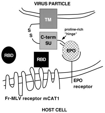
Figure 4.
Schematic diagram of the proposed mechanism of RBD-dependent rescue of Fr-MLV (Epo-env) infection. The viral membrane containing the chimeric Epo-env is at the top. The cellular membrane containing the viral receptor (mCAT1) and the EpoR is at the bottom. Soluble RBD is depicted as a circle that, on receptor contact, undergoes a conformational change (receptor-bound RBD is now a square) that is required for activation of infection through direct interaction with the envelope protein.

