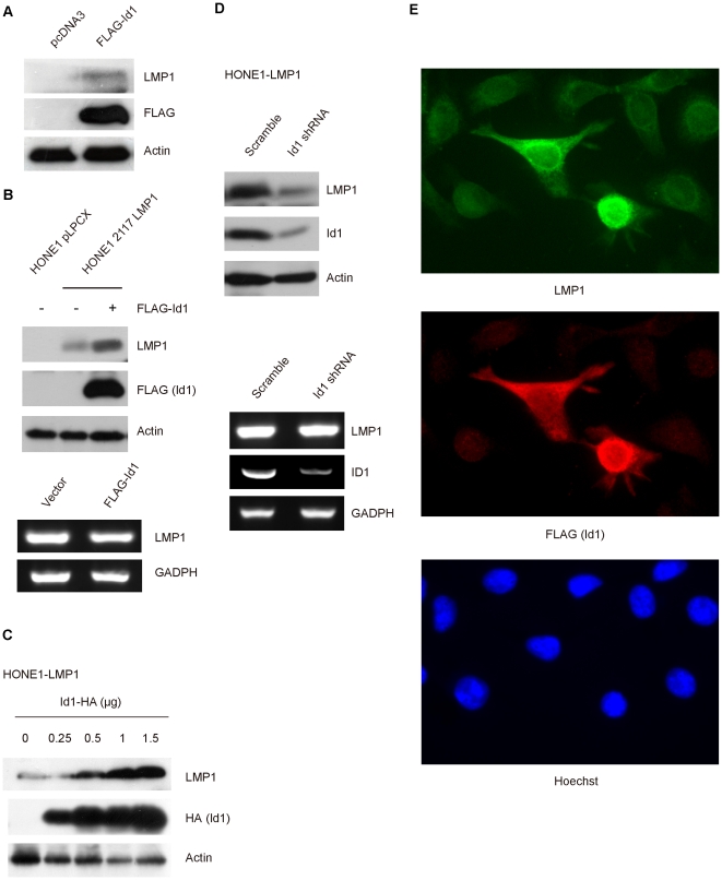
Figure 2. Upregulation of LMP1 level in epithelial cells with Id1 overexpression.
A, The levels of LMP1 protein could be detected after overexpressing Id1 in C666-1 cells. C666-1 cells were transiently transfected either with vector alone or FLAG-Id1, and were lysed 48 hours post-transfection. Protein were transferred to PVDF membrane and subjected to Western blot analysis with anti-LMP1 and anti-FLAG antibody. B, Increase of LMP1 protein levels in HONE1-2117 LMP1 stable cells. HONE1-2117 LMP1 cells were transiently transfected with FLAG-Id1. Cells were examined by Western blot analysis for LMP1 (left panel) and RT-PCR for LMP1 expression with specific primers as described under “Experimental Procedures” (right panel). C, Id1 upregulated LMP1 in a dose-dependent manner. Increasing amount of Id1-HA plasmid was transfected into HONE1-2117 LMP1 which stably expressed LMP1. Western blotting was performed 48 hours post transfection. The amount of plasmids used are indicated on the figure in µg. Protein extracts were subjected to Western blot with anti-LMP1 and anti-HA antibody. Actin was used as loading control. D. Id1 knockdown was followed by decrease of LMP1 protein level. HONE1-2117 LMP1 was transiently transfected with either scramble shRNA or Id1 specific shRNA. Cells lysates were subjected to Western blotting analysis with anti-LMP1 and anti-Id1 antibody (left panel). RT-PCR was performed with specific primers to detect LMP1 and ID1 gene transcripts. GADPH was used as loading control. E. Immunofluorescence examination of HONE1-2117 LMP1 cells transiently transfected with FLAG-Id1. HONE1-2117 LMP1 cells were transfected with FLAG-Id1 plasmid and immunostained with anti-LMP1 (green), anti-FLAG (Id1, Red) antibody and counterstained with Hoechst 33258 to locate the nucleus (see “Experimental Procedures for further details). Higher expression of LMP1 was observed in HONE1-2117 LMP1 cells overexpressing Id1.

