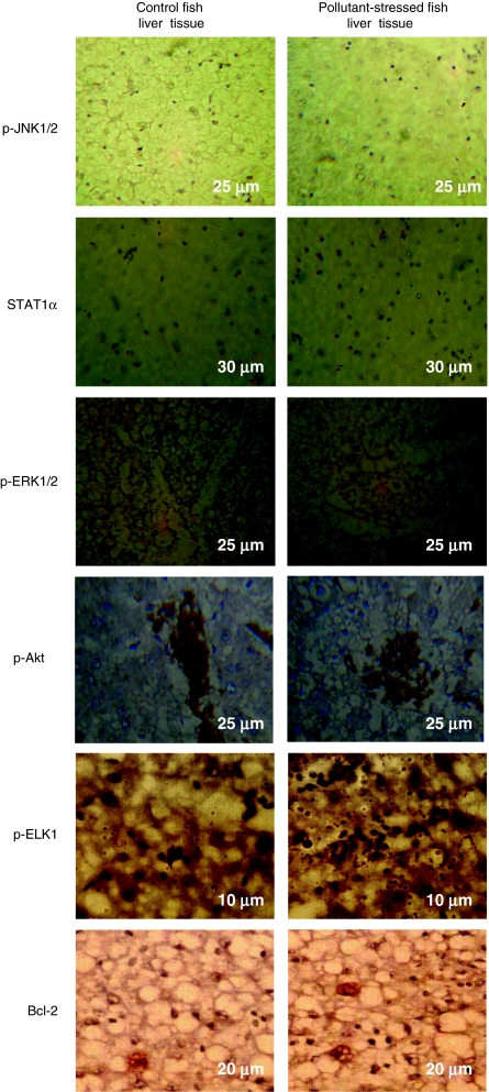
Fig. 4.
Immunohistochemical stainings of signaling proteins like p-JNK1/2, STAT1α, ERK1/2, p-Akt, p-ELK1, and Bcl-2 in the liver tissue of M. cephalus inhabiting the control and pollutant-stressed estuaries. Liver tissue of fish from the control estuary shows light immunostaining, whereas liver tissue of fish from the pollutant-stressed estuary shows intense immunostaining of all signaling molecules except for p-JNK1/2, the results being confirmed from average staining intensity of signaling proteins. The measurements in the micrographs represent scale bar size

