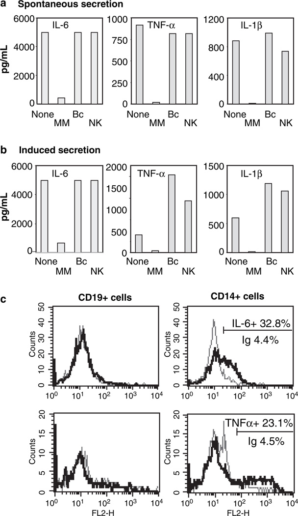
Fig. 2.
Monocytes secrete cytokines both spontaneously and in response to Toll-like receptor stimulation. (a) Untreated peripheral blood mononuclear cells (PBMC) (None) or PBMC depleted from monocytes (CD14+), B cells (CD19+) or NK cells (CD56+) by immune-magnetic separation were cultured in medium alone. (b) PBMC untreated or population-depleted were stimulated with lipopolysaccharide for 16 h. Secreted cytokines in the supernatants were evaluated by cytometric bead array assay. Data shown [from a patient with hepatitis C virus (HCV) mono-infection] is representative of identical results from four separate patients. (c) Intracellular staining for interleukin (IL)-6 (upper row) in B cells (CD19+) and monocytes (CD14+) from a patient with human immunodeficiency virus/HCV co-infection. Staining in NK cells (CD56+) was also negative (data not shown). The grey line depicts the flow cytometric profile with an isotype control antibody (4.4% of CD14+ cells) and the black line indicates IL-6 staining (32.8% of the CD14+ population). Staining for tumour necrosis factor (TNF)-α was positive in 23.1% of CD14+ cells (4.5% for the isotype control). No TNF-α signal was detected in CD56+ (NK) cells (data not shown).

