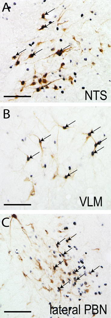
Figure 3. LPS-induced Fos activation of brainstem vlBNST afferents.
LPS treatment activated vlBNST-projecting neurons within the NTS (A), VLM (B), and lateral PBN (C), as evidenced by Fos expression (black nuclear staining) within retrogradely-labeled neurons (brown cytoplasmic labeling). Arrows point out examples of double-labeled neurons. Scale bars = 100 µm.

