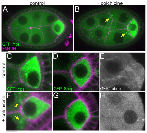
Fig. 4. The localization of GFP::Yps, but not GFP::Shep was affected by colchicine treatment.
Stage-6 egg chambers were incubated in culture medium without (control: A, C–E) or with 50 μg/ml colchicine (B, F–H) for 1 hr. GFP::Yps was enriched in the oocyte (A and C). When MTs were disrupted, GFP::Yps mislocalized in nurse cells and made large aggregates in cytoplasm (arrows in B and F). See also Movie S2. In contrast, the distribution of GFP::Shep was not detectably affected by colchicine (D and G). (E and H) Control and disrupted MTs were visualized by GFP::tubulin. Bars, 10 μm.

