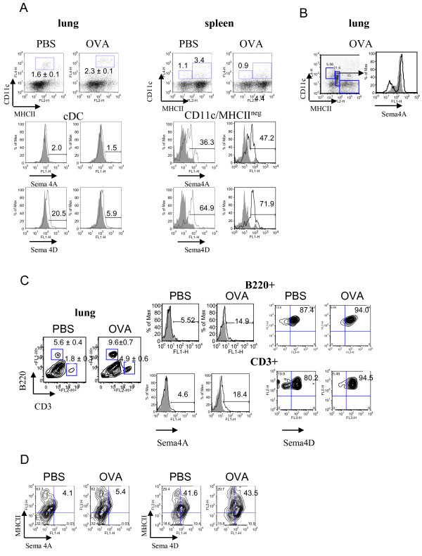
Figure 3.
Regulation of Sema4A and Sema4D expression on lung immune cells by allergen and VEGF. Flow cytometric detection of Sema4A and Sema4D expression on lung and spleen DC (A-B), lung T and B cells (C), and MHCII+ cells (D). Single cell suspensions were prepared as described in Methods. Conventional DC were identified by staining cells with anti-CD11c, -MHCII, and -CD11b Abs used for lung cDC detection. Highly fluorescent macrophages were gated out from the further analysis as large cells on FSC-SSC. (A) No cell surface Sema4A and low Sema4D expression was found on lung cDC. Spleen CD11c+MHCIIneg DC subset demonstrated high Sema4A and Sema4D expression which was further upregulated by allergen. (B) Intracellular Sema4A was targeted to a specific population of CD11cintermed/MHCIIlow cells under inflammatory conditions. (C-D) Low Sema4A and high Sema4D expression was detected on lung B220+ cells, CD3+ cells, and MHCII+ cells.

