Figure 4.
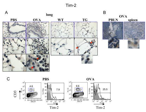
Regulation of lung Tim-2 expression by allergen and VEGF. Immunohistochemical (A-B) and flow cytometric (C) detection of Tim-2 expression in lung (A) and lymphoid (B) tissues, and on lung T cells (C). (A-B) Note positive Tim-2 staining on different lung cells besides lymphocytes in allergen- or VEGF-exposed mouse lungs. Red arrows point to Tim-2+ APC-like cells and granulocyte. Inserts show high magnification fields (100x) with marker-positive cells (lymphocytes). (C) Flow cytometry analysis of cells from lung enzymatic digests obtained from PBS- and allergen-treated mice showed an increase in lung Tim-2+ T cells in OVA-treated mice. The histograms show the percentage of Tim-2+CD3+ cells in the lung (clear histogram) as compared to the appropriate isotype control stained CD3+ cells (gray histogram).

