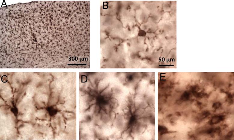
Figure 10.
High-power views of the distribution and morphology of Iba1-immunoreactive cells following 2 closed-skull impacts (TBI-TBI). (A) There are uniformly distributed Iba1-positive microglia in the ipsilateral cortex at 7 days after the first of 2 impacts. (B) Ramified morphology (resting) morphology. (C) Hypertrophic morphology (activated). (D) Bushy morphology (activated). (E) Ameboid morphology (activated). Scale bar (50 = μm) in panel B applies also to panels C-E.

