Figure 7.
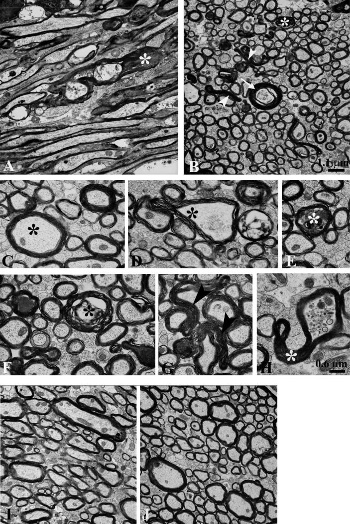
Electron microscopy reveals prominent axonal cytoskeletal injury in 2 closed-skull impacts (TBI-TBI) mice. Images from the ipsilateral external capsule 7 days after the second of 2 impacts. (A) In a section parallel to the predominant direction of axonal fibers individual myelinated axons are dilated and contained intra-axoplasmic electron dense material (*) with cytoskeletal disruption. Many preserved myelinated and unmyelinated axons are also present. (B) A transverse section shows dystrophic myelinated axons (*), axoplasmic collapse (arrowheads), and cytoskeletal disruption (arrow). (C) High magnification cross-sectional view of a normal myelinated axon with intact cytoskeleton (*). (D) Axon with partial axoplasmic collapse and cytoskeletal compaction (*). (E) Dystrophic myelinated axon with intra-axoplasmic abnormal electron dense material (*). (F) Multiple abnormal axons, including double concentric myelin sheaths (*) suggesting possible invagination. (G) Axoplasmic collapse across 2 myelinated axons (arrowheads). (H) Partial axoplasmic collapse (*) and abnormal accumulation of organelles. (I, J) No axonal cytoskeletal abnormalities in the external capsule of control mice prepared for electron microscopy 4 days after the second of 2 sham impacts. Scale bar (1.1 μm) in panel B also applies to panels A, I and J. Scale bar (0.6 μm) in panel H also applies to panels C–G.

