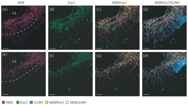
Fig. 2.
Kisspeptin (Kiss1) and neurokinin B (NKB) fibre distribution in the median eminence (ME). NKB-immunoreactive (−ir) (A, E; magenta) and Kiss1-ir (B, F; green) fibres were found in the ME and overlay of these images revealed colocalised Kiss1/NKB-ir fibres (C, G; yellow) primarily in the internal zone. Doublelabelled Kiss1/NKB-ir fibres were observed in close proximity to gonadotrophin-releasing hormone-green fluorescent protein (GnRH-GFP) fibres (D, H; blue). Single-labelled Kiss1-ir fibres were also observed primarily in the internal zone, whereas single-labelled NKB-ir fibres were observed in both the internal and external zone of the ME. Single-labelled NKB-ir fibres were often found in close proximity to GnRH fibres in the external zone of the ME (white). Two ME examples from the same animal are shown to illustrate variability of staining within the external zone. The dotted lines in (A) and (E) mark the approximate border of the internal and external zone. Immunohistochemistry was performed in ovariectomised GnRH-GFP virgin rats. Scale bars = 50 μm; V3, third ventricle.

