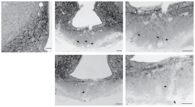
Fig. 3.
Neurokinin B (NKB) immunoreactivity in the arcuate nucleus (ARH) and median eminence (ME) using nickel-intensified 3,3′-diaminobenzidine tetrahydrochloride (NiDAB) secondary detection. NiDAB staining was used to verify the variable quantity of NKB fibres in the external zone of the ME as observed by fluorescence labelling. ARH NKB-immuoreactivity (ir) (A) appeared similar to fluorescence immunoreactivity. The quantity of NKB-ir fibres in the external zone of the ME (arrows) varied from moderate (B, C) to light (D, E) in sections taken from the same ovariectomised virgin rat. (C, E) Magnifications of (B) and (D), respectively. Scale bars = 50 μm.

