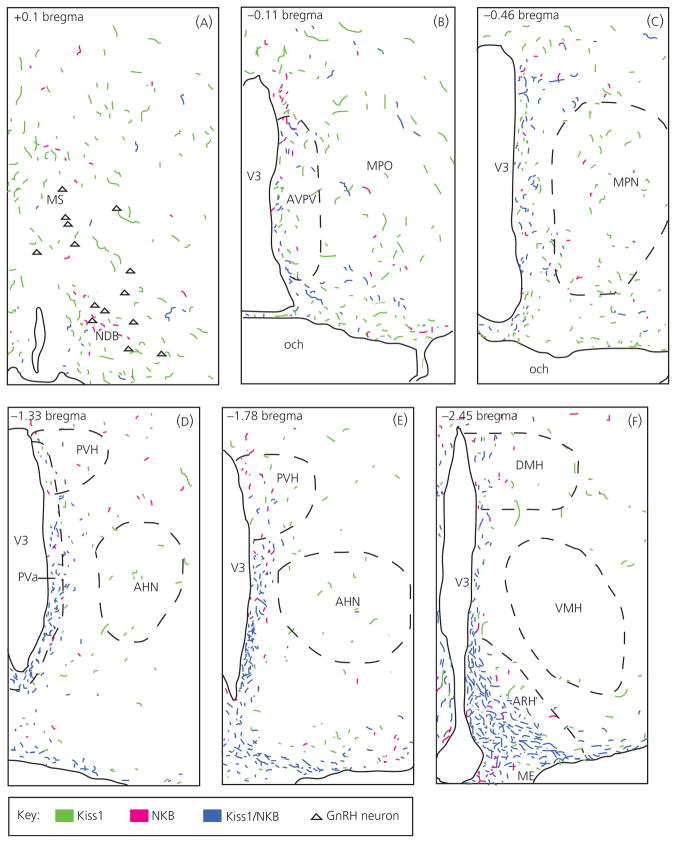
Fig. 4.
Computer assisted line drawings of kisspeptin (Kiss1)/neurokinin B (NKB)-immunoreactive (ir) rostral fibre projections from the arcuate nucleus. Double-labelled Kiss1/NKB-ir fibres (blue) in ovariectomised virgin control tissue had a rostral projection pattern that closely followed the third ventricle (V3) with fibres diminishing rostrally (panels A–F represent a rostral to caudal progression, beginning at the level of the NDB in panel A and ending at the level of the ARH in panel F). Very few double-labelled Kiss1/NKB-ir fibres were observed near gonadtophin-releasing hormone (GnRH) neurones (A; triangles) in the NDB, although single-labelled Kiss1-ir fibres (green) and NKB-ir fibres (magenta) were found in this area. Corresponding coordinates from the Swanson rat brain atlas (30) are given in upper left corner of each panel. AHN, anterior hypothalamic nucleus; AVPV, anteroventral periventricular nucleus; DMH, dorsomedial nucleus of the hypothalamus; ME, median eminence; MPN, medial preoptic nucleus; MPO, medial preoptic area; MS, medial septal nucleus; NDB, nucleus of the diagonal band of Broca; och, optic chiasm; VMH, ventromedial nucleus hypothalamus.

