Figure 3.
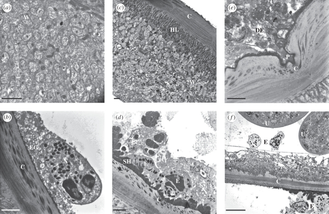
Ultrastructure of O. ochengi onchocercomata at 12 weeks. (a) Hypodermis of an untreated worm showing abundant Wolbachia endosymbionts (W). (b) The cuticle (C) of an untreated worm displaying normal structure, alongside an adherent neutrophil (N) surrounded by numerous extracellular granules (EG). (c) A worm section from the melarsomine (MEL) group displaying intact Wolbachia (W) beneath the hypodermal lamellae (HL) and the cuticle (C). (d) Eosinophils (E) attached to Splendore-Hoeppli deposits (SH) on the cuticle (C) of a worm from the MEL group. (e) The pseudopodium (P) of a degranulating eosinophil (DE) entering a cleft in the cuticle (C) of an oxytetracycline (OXY)-treated worm. (f) Two eosinophils (E) adjacent to the paired uteri (U) within the pseudocoelomic cavity of a worm from the OXY group; eosinophils are also visible on the host side of the cuticular (C) interface. Note the highly vacuolated hypodermis. Scale bars: (a–e) 2 µm; (f)10 µm.

