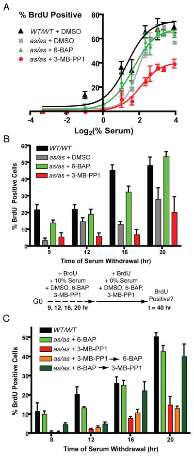
Figure 6. Manipulating Cdk2 with small molecules uncovers a non-redundant role in R-point control.
(A) Wild-type and Cdk2as/as RPE-hTERT cells were synchronized in G0 by contact inhibition and released into medium containing nocodazole, BrdU, the indicated concentration of serum, and DMSO, 0.5 μM 6-BAP or 10 μM 3-MB-PP1. Cells were fixed 40 hr after release and stained to detect BrdU incorporation.
(B) Wild-type and Cdk2as/as RPE-hTERT cells were synchronized in G0 by contact inhibition and released into 10% serum-containing medium supplemented with BrdU and DMSO, 0.5 μM 6-BAP or 10 μM 3-MB-PP1, for 9, 12, 16, or 20 hr. Cells were washed and changed to serum-free medium containing BrdU while maintaining drug treatment. Cells were fixed 40 hr after release and stained to detect BrdU incorporation.
(C) R-point passage was determined as in (B), except that when cells were transferred from 10% to 0% serum, drug treatment was switched from 3-MB-PP1 to 6-BAP (orange bars) or from 6-BAP to 3-MB-PP1 (dark green bars).
Each data point is the mean +/− SEM of 4 fields of cells.

