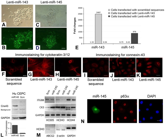
Figure 3. Transfection analysis of miR-143 and miR-145.
(A–D) Human P2 CEPCs transfected with (A and B) Lenti-miR-143 and (C and D) Lenti-miR-145. (A, C) Phase-contrast images; (B, D) live GFP imaging. (E) Overexpression levels of miR-143 and 145 in transfected CEPCs by qPCR analysis. Amplification signals from cells with scrambled sequences (Scm) were indicated. Immunofluorescence of (F–H) cytokeratin-3/12 and (I–K) connexin-43 in CEPCs transfected with (F, I) scrambled sequence, (G, J) Lenti-miR-143 and (H, K). (L) Western blotting and densitometry analysis of connexin-43 (Cnx43) and GAPDH in CEPCs transfected with Lenti-miR-145 or scrambled sequences. (M) RT-PCR result of integrin β8 (ITGB8), cytokeratin-3 (CK3), ABCG2, β-actin and GAPDH in different primary CEPCs (at P2) transfected with Lenti-miR-145 or scrambled sequences. (N) Immunofluorescence of miR-145 (revealed by GFP), p63α and nuclear DAPI stain in P2 CEPCs after Lenti-miR-145 transfection. Scale bars: (A–D) 100 µm; (F–K, N) 10 µm.

