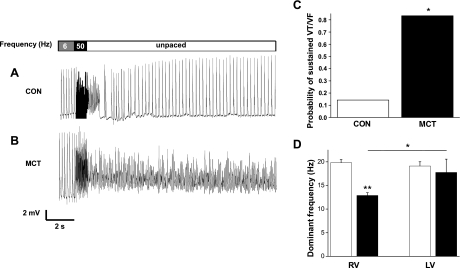
Fig. 4.
Arrhythmias in Langendorff-perfused hearts. MAPs from a CON (A) and MCT (B) heart. Hearts were initially paced at 6 Hz, then a 1-s burst stimulus was applied to induce ventricular tachycardia (VT) or fibrillation (VF), and external stimulation was then stopped. After a brief period of arrhythmia the CON heart recovers a stable, intrinsic rhythm; however, the VF generated in the MCT heart is sustained. C: probability that CON and MCT hearts show sustained VT or VF (*P < 0.05, CON vs. MCT). D: dominant frequency of arrhythmias from CON (open bars) and MCT (closed bars) RV and LV (**P < 0.01, CON RV vs. MCT RV; *P < 0.05, MCT LV vs. MCT RV; n = 7 CON and 6 MCT).

