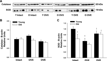
Fig. 7.
A: representative immunoblots of catalase and SOD protein expression in coronary arterioles from young and aged female rats. B: arteriolar catalase expression did not vary with age or estrogen status (n = 7–8) while SOD protein levels declined with both age and OVX. C: protein expression for catalase and SOD was normalized to the β-actin blot shown in Fig. 6A. In coronary microvessels from aged females, SOD expression increased following estrogen treatment (n = 6–8). Data are presented as means ± SE. *P < 0.05 vs. young intact. †P < 0.05 vs. age-matched intact. ‡P < 0.05 vs. age-matched OVX.

