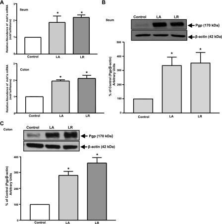
Fig. 4.
P-gp expression in response to L. acidophilus or L. rhamnosus [3 × 10⁹ colony-forming units (CFUs)]. A: relative abundance of mdr1a mRNA from ileum and colon of control and treated mice normalized to histone mRNA (internal control). Total lysates extracted from mucosal tissues of ileum (B) and colon (C) of control and treated mice were subjected to 7% SDS-PAGE and then transferred to a nitrocellulose membrane. Blot was probed with anti-mdr1 (P-gp) or anti-β-actin (β-actin) antibody. A representative blot is shown. Data were quantified by densitometric analysis and are expressed as percentage of control in arbitrary units. Values are means ± SE of 3 mice. *P < 0.05 vs. untreated control.

