Fig. 1.
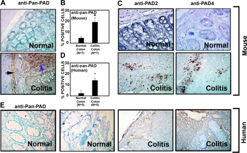
Peptidylarginine deiminase (PAD) levels are elevated in mouse and human colitis. A–C: initial screening for which tissues archived from previous studies using the dextran sulfate sodium (DSS) mouse model of colitis were used (11, 15). Colon sections from 7 mice per group were stained (at the same time) with the polyclonal anti-pan-PAD antibody (A) by rocking using the Antibody Amplifier to ensure even and reproducible staining. Antibody recognizes multiple isozymes of PAD and is not specific to any 1 of the 5 known forms. Importantly, Cl-amidine is also a ubiquitous inhibitor of PAD isozymes. Staining in normal colon (top) was largely absent in the mucosa. In colitis (bottom), staining was apparent not only in inflammatory cells and the lamina submucosa (black arrow), but also in cells of the lamina propria (blue arrow). B: quantification with the use of an automated cellular imaging system. *Significantly increased PAD levels in colitis (P < 0.001). C: antibodies against PAD2 and PAD4 also showed increased levels of these two isozymes in mouse colon colitis tissues. D: quantification of human tissues [colon tissues from colitis patients with diagnostically inflamed colons (n = 5) and normal colon tissues from patients without colitis (n = 5)] by automated cellular imaging system. *Significantly increased PAD levels in colitis (P < 0.01). E: normal human colon tissue and colon tissue from patients with ulcerative colitis probed with the anti-pan-PAD antibody. Staining appears to be specific to inflammatory cells.

