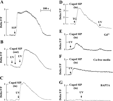
Fig. 1.
Effect of sphingosine-1-phosphate (S1P) and caged S1P on [Ca2+]i. Endothelial cells (ECs) grown on 35-mm glass-bottom dishes were loaded with calcium fluorescent indicator Fluor-4 AM, and intracellular ionic Ca2+ was monitored by confocal microscopy as described in materials and methods. A: ECs were challenged with S1P (1 μM). B: caged S1P (1 μM) was added to the incubation media. C: ECs were preloaded with caged S1P for 15 min, cells were washed, and intracellular calcium was monitored. D: same as C, but cells were treated with 5 μM thapsigargin (TG) before UV flash. E–G: same as C, but cells were pretreated with Ga3+ (1 μM, 15 min) (E), or incubated in Ca-free media (F), or pretreated with BAPTA (25 μM, 1 h) (G) before UV flash. As expected, treatment with TG resulted in the depletion of intracellular Ca2+ stores, and further exposure to UV had no additional effect, demonstrating that intracellular S1P targeted intracellular stored [Ca2+]i. Representative tracings from 3 independent experiments are shown.

