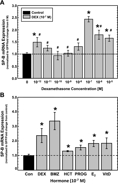
Fig. 4.
Effects of DEX concentration and other steroid hormones on steady-state levels of SP-B mRNA. A: changes in steady-state levels of SP-B mRNA in response to various concentrations of DEX. A549 cells were transfected with pCMVGFP-hspB:N and incubated in the absence or presence of indicated concentrations of DEX. RNA was isolated and subjected to Northern analysis for the presence of SP-B and GFP mRNA, and the SP-B mRNA/GFP mRNA ratio was determined in each sample. The average ratio in untreated samples was set as 1; the ratio from DEX-treated samples was normalized to this average. Shown are normalized SP-B levels in DEX-treated samples relative to levels in untreated samples (means ± SE; N ≥ 7, 2 independent experiments; *P < 0.02 relative to untreated controls; #P < 0.02 relative to levels at 10−7 M DEX). B: changes in steady-state levels of SP-B mRNA in response to other steroid hormones. A549 cells were transfected with pCMVGFP-hspB:N and incubated for 36 h in the absence (Con) or presence of various steroid hormones (10−7 M), including DEX, betamethasone (BMZ), hydrocortisone (HCT), progesterone (PROG), β-estradiol (E2), and vitamin D (VitD). RNA was isolated and subjected to Northern analysis. The levels of SP-B and GFP were quantified, and the SP-B mRNA/GFP mRNA ratio was determined in each sample. The average ratio in untreated samples was set as 1; the ratio from the hormone-treated samples was normalized to this average. Shown are normalized SP-B levels in treated samples relative to levels in untreated samples (means ± SE; N ≥ 7, 2 independent experiments; *P < 0.03 relative to untreated controls).

