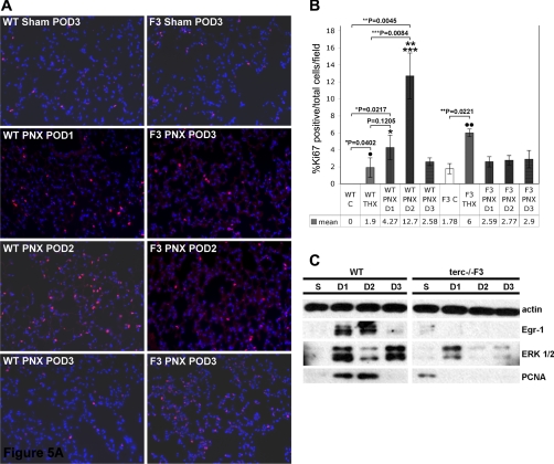
Fig. 5.
Proliferation marker expression in whole lung and AEC2 WT and terc−/−F3 post-PNX. A: Ki-67 expression in whole lung. Representative sections from sham-operated samples at POD3 and postpneumonectomy samples harvested at POD1, POD2, and POD3 (D1, D2, and D3, respectively) were fixed and subjected to immunohistochemistry using Ki-67 primary antibody and Cy-3 secondary antibody (red). Nuclei were stained with DAPI. Nonoperated control samples were also prepared and analyzed for Ki-67 expression (not shown). Negative controls for Ki-67 antibody binding were performed on adjacent sections using mouse IgG in place of specific primary antibody (not shown). B: percentage of Ki-67-positive cells per total cells per field in WT and terc−/−F3 lung post-PNX. For each group, n = 6 (fields chosen from slides containing sections from 2 animals with 3 sections/slide). Ki-67-positive cells per total number of cells per microscopic field were quantitated by calculating the percentage of marker positive (red) nuclei per total nuclei as stained by DAPI in each ×20 field analyzed. The percentage of Ki-67-positive cells in WT POD2 lung was significantly higher than the percentages in WT nonoperated control (**P = 0.0045) and WT THX POD3 control (***P = 0.0084). WT POD1 and POD3 samples exhibited significantly more Ki-67-positive cells than nonoperated control (*P = 0.0217 for PNX POD1), but not sham-operated control (P = 0.1205 for PNX POD1). Both WT and terc−/−F3 THX control samples showed elevated Ki-67 expression compared with nonoperated controls and in both cohorts and these differences were significant by Student's t-test (·P = 0.0217; ··P = 0.0221). No significant change in the percentage of Ki-67-positive cells was observed in post-PNX terc−/−F3 lung samples compared with terc−/−F3 nonoperated control at any post-PNX time point. All post-PNX terc−/−F3 samples exhibited significantly fewer Ki-67-positive cells than sham-operated control samples. C: expression of proliferative markers early growth response protein-1 (Egr-1), MAP kinase ERK1/2, and PCNA in AEC2 harvested post-PNX from WT and terc−/−F3 mice. AEC2 were isolated from right lungs harvested from sham-operated mice at POD3 (S) and from pneumonectomized mice at POD1, POD2, and POD3. Cell lysates were probed for expression of Egr-1, PCNA, and MAP kinase ERK1/2, with expression of actin used as a loading control. Blot shown is representative of multiple blots (3–5) using samples from 3–4 different animals/time point.

