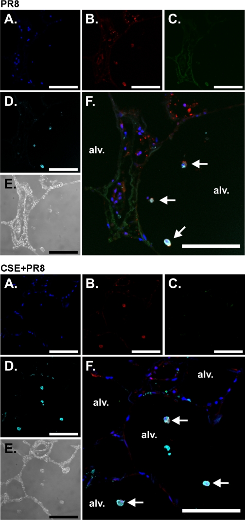
Fig. 7.
CSE suppresses IP-10 induction by influenza virus in macrophages in the human lung. Lung slices were exposed to 6 × 10⁶ PFU/ml of influenza virus PR8 or virus diluents for 24 h in the presence of brefeldin A (BFA) to enhance the detection of cytokines. Slices were then processed for immunohistochemistry for the detection of the chemokine IP-10 using goat polyclonal antibodies, viral nucleoprotein (NP) using rabbit polyclonal antibody, and macrophages using anti-CD68 monoclonal antibody. Nuclei were stained with SYTOX green. Top: PR8 alone. Bottom: CSE + PR8. A–D: fluorescent images that demonstrate nuclei (A; blue), NP (B; red), IP-10 (C; green), and macrophages (D; cyan). E: bright-field images that demonstrate that lung architecture is preserved during the experiment. F: overlays of the fluorescent images that demonstrate that the primary cellular source of IP-10 is alveolar macrophages (arrows). Bars = 100 μm.

