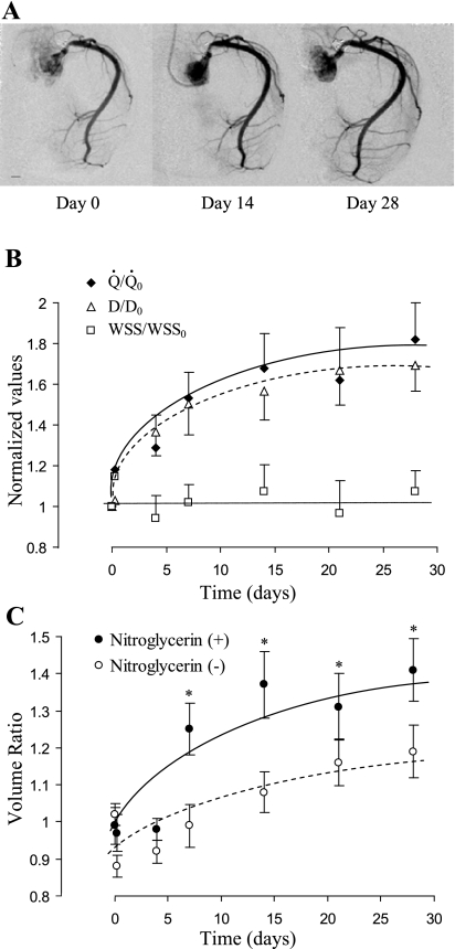
Fig. 2.
A: video-densitometric images of RCA were from the same pig to show the progress (day 0, day 7, and day 28) of RCA remodeling after pulmonary artery (PA) banding. All images are taken at the same magnification. Scale length is 5 mm. B: flow and inner diameter were measurements based on digital subtraction angiography (DSA), and wall shear stress (WSS) was calculated based on the ratio of flow to diameter cubed. Normalized flow (Q̇/Q̇0), diameter cubed [(D/D0)3], and WSS (WSS/WSS0) were defined as the ratio at a given day relative to day 0. Both Q̇/Q̇0 and (D/D0)3 increased gradually with time (one-way ANOVA, P < 0.05). The increase is exponential thereafter, as shown through the best fit line. WSS/WSS0 showed no significant change with time (one-way ANOVA, P > 0.05). C: volumetric growth of RCA from day 0 to day 28. In vivo lumen volume was measured using DSA techniques based on video densitometry. The volume ratio was calculated as the ratio of volume at a particular time point to day 0 (before banding). At the particular time point, lumen volume was first measured without administration of nitroglycerin (basal tone) and then measured again with administration of nitroglycerin as a bolus (0.3 mg, intracoronary) at the inlet of the RCA. The volume ratio of RCA at basal tone significantly increased during RVH (one-way ANOVA, P < 0.05). With administration of nitroglycerin, the volume ratio was significantly increased (two-way ANOVA, *P < 0.05). Data are expressed as means ± SE.

