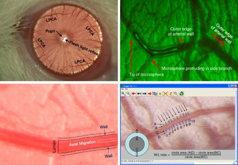
Fig. 1.
Top left: iris of a rat photographed through a slit-lamp biomicroscope (Topcon SL-D7) with a ×40 objective lens (no teleconverter used for this image). The light reflex from the flash light can be seen within the pupil. Four branches of the long posterior ciliary artery (LPCA) can be seen in the superior and inferior and lateral and medial aspects of the iris. The blood vessels projecting in perpendicular direction to the LPCA can be used as anatomical landmarks to identify the same section of the LPCA in subsequent imaging sessions. Top right: identification of the structures representing the vessel wall and lumen was accomplished by intravenous injection of a fluorescent-labeled bolus. The outer edges of the fluorescent-labeled bolus mark the lumen of the blood vessel. Dark lines outside the vessel lumen mark the vessel wall. Bottom left: magnified view of the LPCA. Dark lines outside the vessel lumen mark the outer edges of the vessel wall. A brighter line in the center of the vessel lumen is caused by axial migration of erythrocytes that reflect the flash light more than the translucent plasma in the outer zones of the vessel lumen. Bottom right: determination of wall-to-lumen (W/L) ratio using the Imager software. The outer and inner edges of the vessel wall (points A, B, C, and D) are manually marked using a cross-hair cursor on 10 cross sections of the vessel (indicated by the 10 arrows). For each cross section, the W/L ratio is calculated as the difference of the circle areas determined by the diameters (A D) and (B C) divided by the circle area determined by the diameter (B C). The average of these 10 W/L ratios is used for statistical analysis.

