Figure 2.

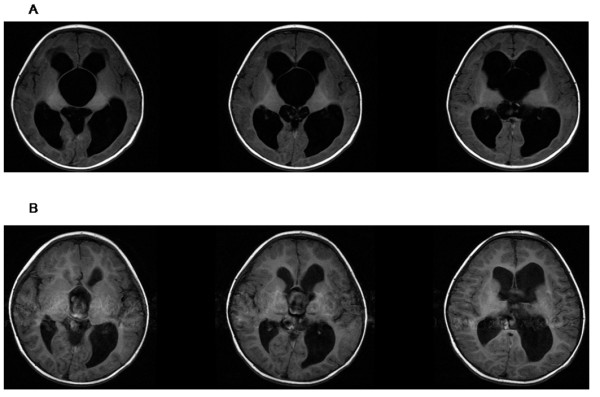
Axial magnetic resonance images obtained in a patient who harbored SSC and hydrocephalus. A: Preoperative axial T1-weighted image (male, 1 year old). B: Postoperative axial T1-weighted image (25 months later).

Axial magnetic resonance images obtained in a patient who harbored SSC and hydrocephalus. A: Preoperative axial T1-weighted image (male, 1 year old). B: Postoperative axial T1-weighted image (25 months later).