Figure 1.
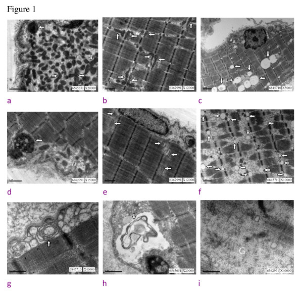
Summarized muscle ultrastructural findings of the four cerebrotendinous xanthomatosis cases. 1a. subsacrolemmal accumulation of mitochondria (arrow), (bar = 1 μm); 1b. mitochondria change in shape and enlarged in size (arrow), (bar = 1 μm); 1c. increased amount of lipid droplets (arrow), (bar = 2 μm); 1d. lipofuscin (arrow), (bar = 1 μm); 1e. triads proliferation (arrow), (bar = 1 μm); 1f. swollen sacroplasmic reticulum (arrow), (bar = 1 μm); 1g. laminated bodies (arrow), (bar = 0.5 μm); 1 h. membranous-like debris (arrow) in the subsacrolemmal space, (bar = 1 μm); 1i. increased amount of glycogen (marked as G, bar = 0.5 μm).

