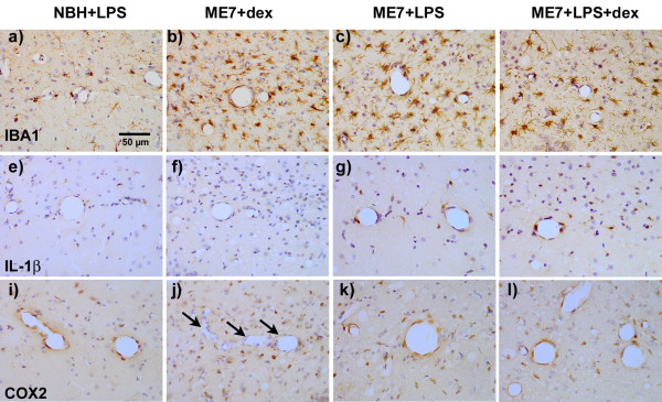
Figure 4.
Immunohistochemical detection of IBA-1, IL-1β and COX2 post-LPS. a-d) IBA-1 labelling of microglia in normal (NBH) and ME7 animals treated with LPS (500 μg/kg) in the presence or absence of dexamethasone-21-phosphate (2 mg/kg) showing increased numbers and activated morphology in all ME7 groups. e-h) IL-1β labelling of microglial cells under the same conditions, showing robust cellular staining for IL-1β only in the ME7+LPS groups, in the presence or absence of dexamethasone. i-l) COX2 labelling of microglial and vascular endothelial cells in the same animals, showing induction of COX2 associated with the vasculature in all animals treated with LPS, but vessels remain unlabelled in ME7 animals treated only with dexamethasone-21-phosphate (arrows). Microglial cells are positively labelled with anti-COX2 antibodies in all groups, but these are more numerous in the ME7 animals. Magnification ×40, Scale bar = 50 μm in all photomicrographs.

