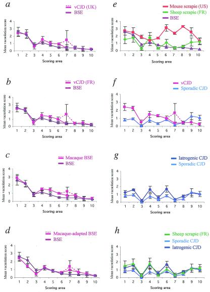
Figure 2.
Lesion profiles in C57BL/6 mice after transmission of vCJD, BSE, sCJD, iCJD, and scrapie. Lesion profiles are (a) cattle BSE and British vCJD (two pooled cases); (b) cattle BSE and the first of the three French vCJD cases; (c) cattle BSE and BSE after primary transmission to macaques; (d) cattle BSE and BSE after secondary transmission to macaques; (e) cattle BSE, the mouse-adapted C506 M3 scrapie strain (derived from U.S. sheep scrapie), natural sheep scrapie (two pooled cases from a single French flock); (f) French vCJD and a French case of sCJD; (g) the former sCJD and one French case of iCJD linked to the administration of growth hormone; and (h) same as in g plus the natural sheep scrapie. The dotted lines belong to the lesion profiles of vCJD (in a and b) and of macaque-BSE (in c and d). Vacuolation was scored on a scale of 0–5 in the following brain areas: 1, medulla; 2, pons/mesencephalon; 3, cerebellar cortex; 4, colliculi; 5, hypothalamus; 6, thalamus; 7, hippocampus; 8, septum; 9, cerebral cortex; and 10, striatum.

