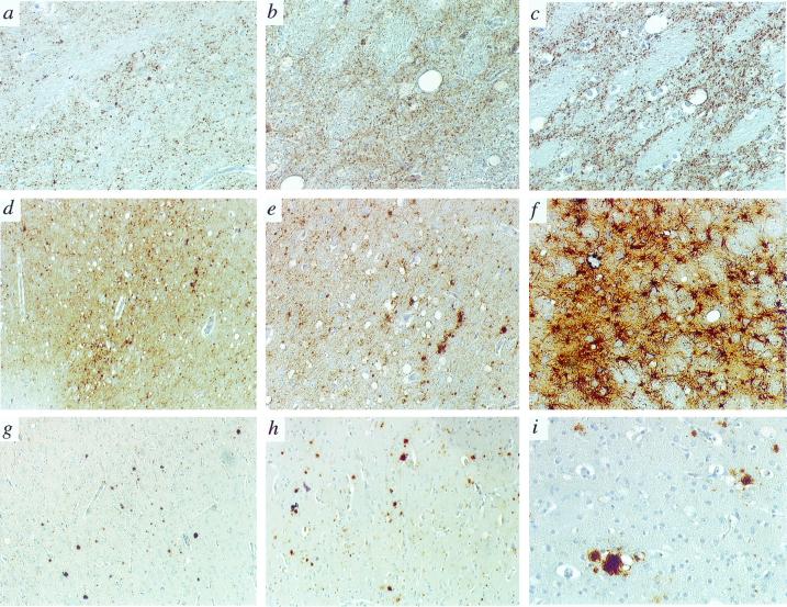
Figure 4.
Immunopathology of BSE, vCJD, and kuru in cynomolgus macaques. All panels show the pattern of PrP deposition with the 3F4 antibody, except f, which depicts glial fibrillary acidic protein immunohistochemistry. (a, b, and c) Thalamus in kuru, BSE, and vCJD, ×20. (d and e) Cerebral cortex in kuru, ×2.5 and ×10. (f) Thalamus in BSE, ×10. (g) Cerebral cortex in BSE (i.v.), ×2.5. (h) Cerebral cortex in BSE (i.c.), ×2.5. (i) Immature florid plaque with a dense core of PrP surrounded by few vacuoles in the cerebral cortex (BSE, i.c.), ×20.

