Fig. 1.
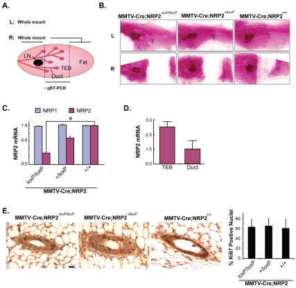
Loss of NRP2 retards mammary gland development. (A) Schematic of mouse mammary gland preparation. Left (L) and right (R) sides of the fourth mammary glands were prepared for each mouse. Extracts prepared from the central area of the right mammary gland were used for qRT-PCR to verify loss of NRP2. The remaining parts of these mammary glands were whole-mounted as shown in B. LN, central lymph node; TEB, terminal end bud. (B) Whole mounts (carmine staining) of 5-week-old virgin mouse mammary glands. Top and bottom pictures show left (L) and right (R) side of the fourth mammary gland. (C) NRP1 and NRP2 mRNA expression in the different genotypes assessed by qPCR. Data were normalized to GAPDH for each sample and expressed as mean±s.d. *P<0.01. (D) Relative NRP2 expression in TEB and mature ducts microdissected from normal mouse mammary glands. Data were normalized to GAPDH for each sample and expressed as mean±s.d. (E) Cell proliferation in mouse mammary glands from 5-week-old MMTV-Cre;NRP2loxP/loxP, MMTV-Cre;NRP2+/loxP and MMTV-Cre mice was compared using Ki67 staining. A minimum of five mice and 30 glands were examined for each genotype. Quantification of Ki67-positive nuclei is shown in the accompanying graph. Error bars represent s.d. Scale bar: 10 μm.

