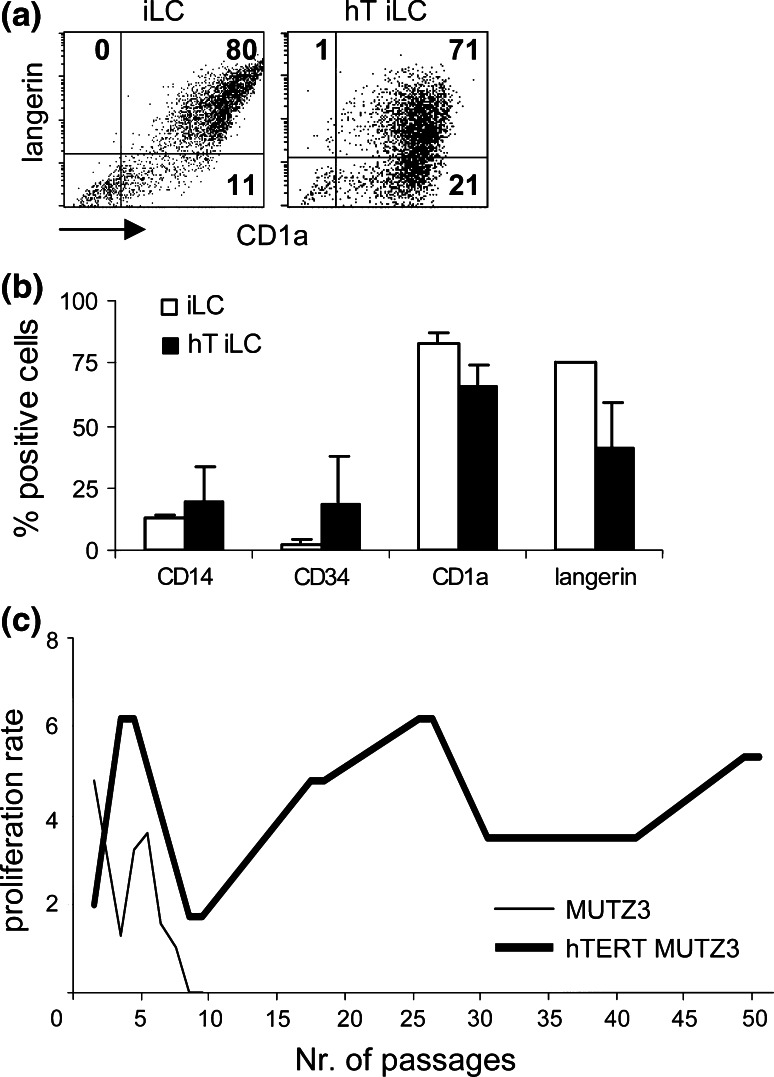
Fig. 2.
Effects of hTERT introduction on MUTZ3-LC differentiation and proliferation. a Dot plots of flow cytometric analysis for the LC-typifying markers CD1a and Langerin on immature LC (iLC) cultures from MUTZ3 (iLC; left) and hTERT-MUTZ3 (hT-iLC; right). b Average percentages (+standard deviation) of CD14+, CD34+, CD1a+ and Langerin+ cells within iLC cultures from MUTZ3 or hTERT-MUTZ3 cells (n = 3). c Progenitor cell proliferation in the presence of 30 nM doxorubicin. Shown are the passage numbers against the expansion of the progenitor cells. MUTZ3 progenitor cells stopped proliferating after 9 passages in the presence of 30 nM doxorubicin, whereas hTERT-MUTZ3 cells could be cultured for over 160 passages (shown are data up to passage 50)

