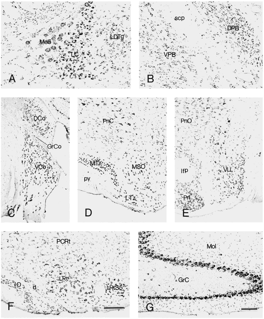
Fig. 11.
CRF1-like immunoreactive neurons in locus coeruleus (A) and parabrachial nuclei (B). C–E: Distribution of labeled neurons in cochlear nuclei (C), trapezoid body (D), and ventral nucleus of lateral lemniscus (E). F,G: Immunoreactive neurons in the inferior olive and cerebellum, respectively. Within the cerebellum, Purkinje cells are intensely labeled. The granular layer contains scattered intensely labeled neurons, as well as numerous weakly stained fine puncta. Scale bars = 200 µm in F (applies to C–F);80 µm in G (applies to A,B,G).

