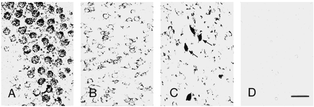
Fig. 2.
Illustration of the three principal patterns of neuronal CRF1-like immunoreactivity (CRF1-LI) in mouse brain. CRF1-LI is characterized as granular (A, cerebellar Purkinje cells), punctate (B, parafascicular thalamic nucleus), or homogenous (arrows in C, lateral hypothalamic area). D: Preadsorption of the antiserum with the immunogenic epitope eliminates all the reaction signal (section shown is from thalamus). Scale bar = 40 µm (applies to A–D).

