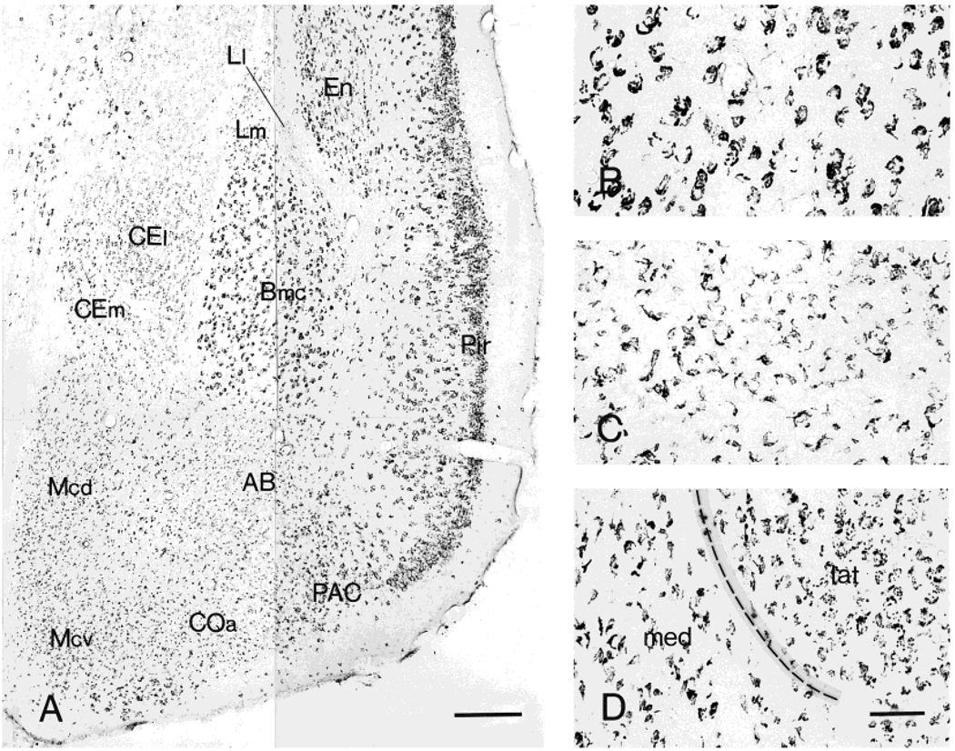
Fig. 5.
Distribution of CRF1-like immunoreactivity in the amygdala (A) and the morphology of neurons showing CRF1-LI in basal (B), medial (C), and central (D) nuclei. These neurons are intensely labeled throughout the magnocellular division of basal nucleus (Bmc). Within the medial nucleus (Mcd and Mcv), CRF1 immunoreaction product frequently outlines only a portion of the cell surface. In the central nucleus, the lateral (CEl) and medial (CEm) divisions show a different staining pattern: whereas immunoreactivity in CEl consists of punctate deposits, the CEm consists of an outline of only a portion of the cell surface. Scale bars = 200 µm in A; 40 µm in D (applies to B–D).

