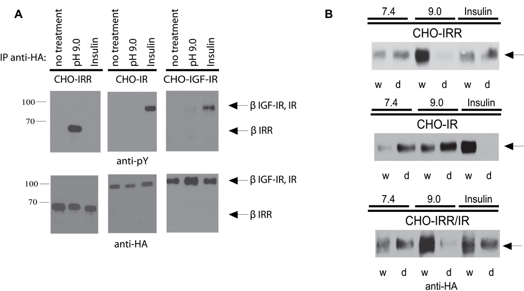
Figure 3. Specificity and structural response to IRR activation by alkali.
(A) Activation of IRR but not of IR and IGF-IR by alkali. After starvation, the indicated stably transfected cell lines were incubated for 10 min in PBS adjusted to pH 9.0 or with 1 µM insulin. Cells were then lysed, immunoprecipitated with anti-HA antibody, and Western blotted with anti-pY or anti-HA antibody. (B) Analysis of IRR, IR and IRR/IR chimera conformational changes by the detergent phase distribution assay. Non-starved stably transfected CHO cells were lysed in Triton X-114 containing buffers adjusted to pH 7.4 or 9.0, or supplemented with 0.5 µM insulin, and analyzed by detergent phase distribution (Florke et al., 2001), as described in detail in Supplemental Experimental Procedures. Equal aliquots of the separated phases (w – detergent-depleted, d – detergent enriched) were Western blotted with anti-HA antibody to determine receptor concentration. See also Figure S2.

