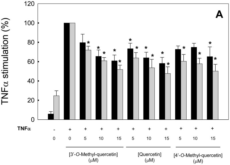
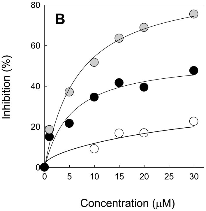
Figure 1. Dose-response effects of 3′-O-methyl-quercetin and 4′-O-methyl-quercetin compared to quercetin on adhesion molecule expression in human aortic endothelial cells.
HAEC were incubated for 18 h in the absence (0 μM) or presence of 5–15 μM quercetin, 3′-O-methyl-quercetin or 4′-O-methyl-quercetin followed by the addition of 100 U/ml TNFα and incubation for 7 h. Panel A shows protein levels of E-selectin (black bars) and ICAM-1 (gray bars) as % of protein levels in HAEC incubated with TNFα in the absence of flavonoids. Results from control cells incubated in the absence of flavonoids and TNFα are also shown. Panel B shows % inhibition of E-selectin (black circles), ICAM-1 (gray circles) and MCP-1 (open circles) expression by 3′-O-methyl-quercetin. Data shown are means ± SEM of at least three independent experiments. One-way ANOVA was used to analyze dose-response trends; *significantly different from 0 μM (Tukey-Kramer, post-hoc analysis).


