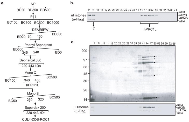
Figure 5.
Purification of a previously unidentified histone ubiquitin E3 ligase complex. a. Schematic representation of the steps used to purify the histone ubiquitin E3 ligase complex. Numbers represent the salt concentrations (mM) at which the E3 ligase activity elutes from the columns. b. Histone ubiquitin ligase assay of protein fractions derived from a Mono Q column. In addition to the ubiquitin ligase activity specific for histone H2A (hPRC1L), a previously unidentified ubiquitin ligase activity for all the core histones was observed in the flowthrough (Ft). c. Silver staining of a polyacrylamide-SDS gel (top) and histone ubiquitin ligase activity (bottom) of fractions derived from a Mono S column. The protein bands that cofractionated with the histone ubiquitin E3 ligase activity are indicated by an asterisk (*). The protein size marker is indicated on the left side of the panel.

